Premature Birth Reshapes Cerebellum Development, Study Finds
A landmark study reveals premature birth disrupts the cerebellum's developmental timeline, linking reduced brain volume to later childhood challenges in movement, language and thinking, challenging the long-held 'catch-up' assumption.
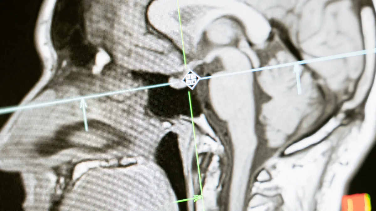
A new study overturns the decades-old belief that premature infants catch up to full-term peers, revealing lasting structural and molecular differences in the cerebellum. Each week spent outside the womb before full term was associated with about 230 cubic millimeters less cerebellar volume, roughly the size of a pea. Between 24 and 40 weeks of pregnancy, the human cerebellum undergoes its most rapid period of development, expanding fivefold in size as its primary neurons wire together.
How premature birth disrupts that growth
Premature birth alters the cerebellum's developmental clock, changing how it grows and develops compared to full-term infants. The entire growth period of the cerebellum happens inside the protective environment of the uterus for a full-term infant. When a baby is born prematurely, this rapid brain construction phase is forced to happen outside the womb.
The analysis revealed that premature birth reshapes the cerebellum's developmental trajectory. The timing of birth influences how distinct brain cell populations mature even after infants reach the same postmenstrual age as those born at term. Spatial transcriptomics technology mapped exact gene activity across intact tissue layers, revealing how major cerebellar neurons-Purkinje and granule cells-develop outside the womb compared with normal gestation. Premature development involves disrupted coordination between these cells.
Molecular markers and long-term outcomes
The study connects the molecular programs set at birth directly to how children grow and develop years later. At 9 to 36 months of age, smaller cerebellar volume correlated with delays in movement, language and thinking skills. The identified molecular markers will improve risk stratification, advance maturity biomarkers and optimize the timing of neuroprotective therapies.
Although birth timing sets the baseline molecular program, physiological stressors, illness and specific aspects of care in the neonatal intensive care unit significantly altered cerebellar growth rates. The molecular programs governing Purkinje and granule cells respond not only to the timing of birth, but also to the clinical environment after birth. The disruption potentially impairs the refinement of neural connections despite the cerebellum retaining its overall structure.
Rethinking assessment and care
The findings necessitate a shift in how preterm infants are assessed and treated. Senior author Panagiotis Kratimenos, a physician-scientist and neonatologist at Children's National Hospital, highlighted the breakthrough. "We could always see the developmental differences that premature children faced, but we could not see the cellular machinery driving them," he said.
Joint senior author Vittorio Gallo emphasized the new guiding principle. "Assessment and treatment of preterm infants should be guided by developmental maturity rather than chronological age," he stated. The study combined multiyear brain imaging and developmental tracking of 176 living infants with cellular analysis of preexisting brain tissue from 83 preterm and term human infants. Samples came from tissue repositories, including the NIH NeuroBioBank.
The research offers a framework for understanding how early birth influences later motor and cognitive outcomes. Although the preterm infant may follow two calendars, one for cerebellar physical growth and one for cellular maturation, neither captures the true complexity of brain health at the time, nor what it means for the child years later.